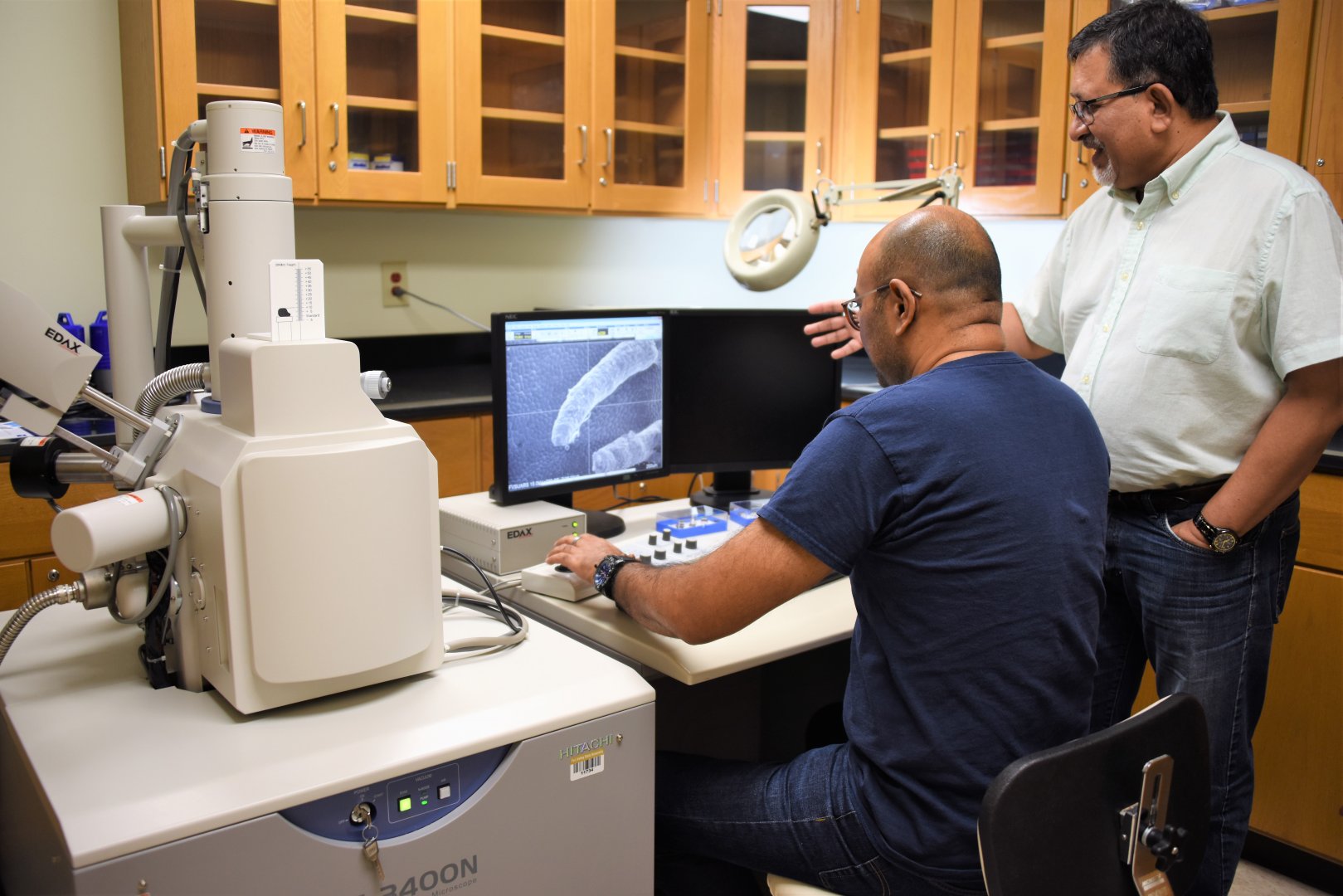
Biological and physical sciences heavily depend on imaging to see beyond the naked eye. An advanced imaging facility at Fort Valley State University is giving graduate students and faculty members the vision to look deeper at issues affecting agriculture and food production.
To further this effort, Dr. Nirmal Joshee, an FVSU plant science professor, assisted Dr. Govind Kannan, FVSU’s associate dean for research, in establishing the Center for Ultrastructure Research (CURE). The new facility, located in the Stallworth Agricultural Research Building, will enhance research capacity, student training and industry partnerships. As the center director, Joshee will also pursue revenue-generating opportunities for the Research Station by allowing other institutions in the state to access the center for a predetermined fee.
“This center will be the only one of its type in middle and south Georgia with both scanning electron microscopy (SEM) and transmission electron microscopy (TEM) capabilities,” Joshee said. “It will be a great selling point in attracting STEM (science, technology, engineering and mathematics) students.”
Electron microscopes allow scientists to obtain images of biological and non-biological specimens at a much higher resolution and magnification (up to 300,000 times) than light microscopes. FVSU’s CURE is now home to these high-quality instruments.
Joshee employed this technology while pursuing his doctorate research from 1981-1986 in India and during his post-doctoral research on cancer cells from 1998-2001 at the University of Nebraska Medical Center in Omaha. After taking a research professional position in 2001 at FVSU, his fascination with electron microscopes led him to bring this advancement to the College of Agriculture, Family Sciences and Technology in 2010.
“Imaging is an important part of science,” he said. “When our students prepare for medical careers in dentistry, ophthalmology or as a researcher, they need imaging concepts. Therefore, we should catch our students early, especially in agriculture.”
Joshee, a medicinal plant researcher, said the SEM and TEM microscopes give a different dimension to research. SEM displays the surface structure of a specimen, whereas TEM shows the internal structure. This requires cutting sections of a specimen and obtaining details at more than one million magnification. Unlike light microscopes that have certain limitations, electron microscopes do not use any lenses for image generation. They use electron beams.
Electron beams hit a study material at a high speed for image generation. To prevent any possible damage, a sample has to go through elaborate processing. These steps include fixation, dehydration, critical point drying and then coating with a thin layer of gold using a sputter coater. As a result, researchers obtain sharp, detailed images of a specimen, which could lead to better research.
The plant science professor said FVSU is already experiencing the reach of this technology. So far, the SEM lab has logged more than 2,000 hours in sample preparation and picture generation for faculty members’ research projects, graduate students’ thesis research and collaborators across the nation.
Brajesh Vaidya, an FVSU research assistant, benefited from using the advanced imaging for his thesis project on Scutellaria, a medicinal plant. He now helps manage the center labs.
The 2012 FVSU Master of Science in biotechnology graduate said he is thankful that the university has this facility on campus for students. “We let them do their own processing,” he said. “We are making them more marketable when they graduate. It always helps to have that extra feather in their hat.”
Scientific instruments worth approximately $1 million are currently in place. Joshee plans to add a small class or demonstration room with audiovisuals and a refurbished walk-in cooler.
For more information about the FVSU CURE, contact Joshee at (478) 822-7039 or josheen@fvsu.edu.

